¹Ð¸²ºÏ CI (±¸, ¸í¹®ÀÇÇм­Àû)
 
 
ÀÇÇм­Àû ȸ¿ø ·Î±×ÀÎ
¾ÆÀ̵ð ÀúÀå
ÀÇÇм­Àû °Ë»ö
 
ÀÇÇм­Àû Ä«Å×°í¸® ã±â
ÀÇÇм­Àû ºü¸¥ ¸Þ´º : ´õ¿í ºü¸£°í Æí¸®ÇÑ ¼­ºñ½º
¸Þ´º¾ó
USMLE
µµ¼­¹®ÀÇ°Ô½ÃÆÇ
¿Ü±¹µµ¼­ ¼öÀÔ½Åû
¿Ü±¹ ÀÇÇм­Àû ÃâÆǻ纰 / ÀÇÇм­Àû °úº°µµ¼­¸ñ·Ï
¾Æ¸¶Á¸ ÀÇÇм­Àû ¹Ù·Î°¡±â
01. ÀÇÇм­Àû¿¡ À¯¿ëÇÑ »çÀÌÆ®
02. ÀÇÇм­Àû ´º½º¸µÅ©
03. ȯÀ²Á¤º¸
Ä«µå»ç¿ë³»¿ª È®ÀÎ : Ä«µå¹øÈ£¿Í »ç¿ë±â°£À» ÀÔ·ÂÇÏ½Ã¸é °Å·¡³»¿ªÀ» Á¶È¸ÇÏ½Ç ¼ö ÀÖ½À´Ï´Ù.
ÀÇÇм­Àû ´Üü±¸¸Å¾È³» : ÀÇÇм­Àû ´Üü±¸¸Å ¹× ÀÇÇм­Àû ´ë·®±¸¸Å¸¦ ¹®ÀÇÇϽøé Ä£ÀýÇÏ°Ô ÇØ°áÇص帳´Ï´Ù.
ÀÇÇм­Àû ±¸ÀÔ ¹«ÅëÀå ÀԱݾȳ» : ¿ì¸®ÀºÇà > 203-05-024131 / ³óÇù > 928-12-030605 | ¿¹±ÝÁÖ : À±ÁøÈñ
±¹¿Ü ÀÇÇм­Àû °Ë»ö
¾Æ¸¶Á¸ ÀÇÇм­Àû °Ë»ö
Barns&Noble ÀÇÇм­Àû °Ë»ö
Home °úº°µµ¼­¸ñ·ÏÀÇÇаúºÐ·ù¸¶ÃëÅëÁõÀÇÇаúAnesthesiology and Pain Medicine
 
 
Atlas of Sonoanatomy for Regional Anesthesia and Pain Medicine, 1st Edition
 

zoom
 
ÃâÆÇ»ç McGraw-Hill Education
ÀúÀÚ Manoj Karmakar
Ãâ°£ÀÏ 2017/12
ÆäÀÌÁö ¼ö 304
Illustrations  
ISBN 9780071789349¡¡(0071789340)
ÆǺ» 1 ÆÇ
Ä¿¹ö Hardcover
ÁÖ¹®¼ö·®
List Price £¤ 155.00
Á¤°¡ £Ü 235,000
ȸ¿ø°¡ £Ü 235,000
¡ØÇØ¿ÜÁÖ¹® µµ¼­ ¾È³» :

- ÇØ¿Ü ¹× ±¹³» ÀÔ°í »çÇ׿¡ µû¶ó 2~8ÁÖ ÀÌ»ó ¼Ò¿äµÉ ¼ö ÀÖÀ¸¸ç. Ãë¼Ò ¹× ¹ÝÇ°ÀÌ ºÒ°¡´ÉÇÕ´Ï´Ù.
- ÇØ¿ÜÁÖ¹®µµ¼­´Â ÇØ¿Ü ÃâÆÇ»ç »çÁ¤¿¡ ÀÇÇØ Ç°Àý/Áö¿¬µÉ¼ö ÀÖ½À´Ï´Ù.
¡Ø±³È¯ ¹× ¹ÝÇ°¾È³» :

- ±³È¯À̳ª ¹ÝÇ°½Ã »óÇ° ¼ö·ÉÈÄ 3ÀÏÀ̳» ±³È¯À̳ª ¹ÝÇ° Àǻ縦 ¾Ë·ÁÁֽðí ÀÏÁÖÀÏÀ̳»¿¡ ÀúÈñÂÊÀ¸·Î »óÇ°ÀÌ ÀÔ°íµÇ¾î¾ßÇÕ´Ï´Ù.
  ½ÃÀÏÀÌ °æ°úµÇ¸é ¹ÝÇ°ÀÌ ºÒ°¡ÇÕ´Ï´Ù.
- º¯½É¿¡ ÀÇÇÑ ±³È¯À̳ª ¹ÝÇ°½Ã, ¹è¼Ûºñ´Â °í°´´Ô²²¼­ ºÎ´ãÇϽøç, ¿À¹è¼ÛÀ̳ª »óÇ° ºÒ·®½Ã¿¡´Â ¹«·á ±³È¯ÀÌ °¡´ÉÇÕ´Ï´Ù.
- ¹ÝÇ°½Ã¿¡´Â ¹ÝÇ°»óÇ° °Ë¼öÈÄ Ä«µå Ãë¼Ò¸¦ Çص帮°Å³ª ÅëÀå,Àû¸³±ÝµîÀ¸·Î ȯºÒ Á¶Ä¡ ÇØ µå¸³´Ï´Ù.
¡Ø¹è¼Û¾È³» :

- Á¦ÁÖµµ¹× µµ¼­»ê°£Áö¿ªÀº Åùèºñ°¡ Ãß°¡µÉ¼ö ÀÖ½À´Ï´Ù.
µµ¼­ ¼Ò°³ | ¸ñÂ÷ | ¼­Æò¾²±â


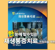
¡¡A comprehensive full-color anatomical atlas designed specifically for the anesthesiologist and pain physician
A clear understanding of relevant anatomy is essential for physicians who wish to master ultrasound-guided nerve blocks. Although ultrasound images may help visualize the nerve to be blocked, the images still present a grainy and incomplete picture. Physicians looking to master US-guided nerve blocks are best served by becoming anatomical experts - and this innovative resource is an invaluable learning tool to doing just that.

In addition to 2D and 3D ultrasound images, Atlas of Sonoanatomy for Regional Anesthesia and Pain Medicine includes high-resolution CTs, MRIs, cadaver anatomy images, and anatomical illustrations to give physicians a comprehensive understanding of the anatomy of the neck, upper and lower extremity, trunk, thorax, thoracic spine, sacral spine, lumbar paravertebral region, and thoracic paravertebral region that are relevant to ultrasound-guided regional anesthesia.

Features
¡¡• Bulleted pearls impart how to obtain optimal ultrasound images at each site
¡¡• More than 600 full-color photographs and illustrations throughout
¡¡• Essential for anesthesiologists and pain physicians, and is also of value to musculoskeletal sonographers, radiologists, and emergency medicine physicians
¡¡• Enables you to learn and see, through full-color illustrations, the relevant anatomy for administering successful nerve blocks
¡¡• Includes CT and MRI correlations to ultrasound images, plus corresponding anatomical illustrations, cadaver anatomy, and photos of probe placement on the patient.

Although other texts may provide some of the imaging information contained in this unique atlas, this is the first resource to systematically and comprehensively gather all the imaging modalities for side-by-side comparison under one cover.


Section I. Neck and Upper Extremity
¡¡1. Exit foramen of the cervical nerve roots
¡¡2. Interscalene groove
¡¡3. Stellate ganglion block anatomy - right and left
¡¡4. Supraclavicular Fossa
¡¡5. Infraclavicular Fossa below the Mid-clavicular point
¡¡6. Infraclavicular Fossa Para-coracoid process
¡¡7. Axilla
¡¡8. Mid-humeral
¡¡9. Arm posterior and lateral aspect for Radial nerve
¡¡10. Elbow – Anterior
¡¡11. Elbow – Posterior for Ulnar nerve
¡¡12. Mid-forearm Scan (Anterior Aspect) for Median, Ulnar and Radial nerves
¡¡13. Panoramic scan of forearm (mid-forearm)
¡¡14. Panoramic scan of the Arm (middle of arm)
¡¡15. Panoramic scan of the mid-forearm
¡¡16. Panoramic scan of the Arm (mid-arm)

Section II. Lumbar Paravertebral Region
¡¡17. Lumbar Paravertebral Region - Right
¡¡18. Lumbar Paravertebral Region - Left

Section III. Lower Extremity with volunteer in the Left Lateral Position
¡¡19. Gluteal Region: Sciatic nerve in the para-sacral area
¡¡20. Gluteal Region: Sciatic nerve in the subgluteal space

Chapter IV. Lower Extremity with volunteer in the Prone Position
¡¡21. Sciatic Nerve in the Infragluteal region
¡¡22. Sciatic Nerve in the mid-thigh posteriorly
¡¡23. Sciatic Nerve in the apex of the popliteal fossa
¡¡24. Sciatic Nerve in the popliteal fossa

Section V. Lower Extremity with volunteer in the Supine Position
¡¡25. Groin t the level of the inguinal ligament – femoral nerve, vein and artery
¡¡26. Groin at the lateral aspect of the inguinal ligament for the Lateral femoral cutaneous nerve
¡¡27. Groin on the medial aspect of the thigh for the Obturator nerve
¡¡28. Mid-thigh scan at the level of the adductor canal
¡¡29. Ankle scan – Medial aspect for the posterior tibial nerve and saphenous nerve
¡¡30. Ankle scan – Lateral aspect for the sural nerve
¡¡31. Ankle scan – Anterior aspect for the deep peroneal nerve and superficial peroneal nerve

Section VI. Sacral Spine
¡¡32. Sacral Hiatus
¡¡33. Dorsal surface of the sacrum
¡¡34. L5Si Gap – lumbosacral junction

Section VII. Lumbar Spine
¡¡35. Lumbar Spine Transverse Scan (L345) – Spinous Process view
¡¡36. Lumbar Spine Transverse Scan (L345) – Interspinous Process view
¡¡37. Lumbar Spine Median Sagittal Scan (L345) – Spinous process view
¡¡38. Lumbar Spine Paramedian Sagittal Scan (L345) – Lamina view
¡¡39. Lumbar Spine Paramedian oblique sagittal Scan (L345) – Lamina view
¡¡40. Lumbar Spine Paramedian Sagittal Scan (L345) – Transverse process view

Section VIII. Thoracic Spine
¡¡41. Upper Thoracic Spine (T1-T4 level)
¡¡42. Mid-thoracic Spine (T5-T8)
¡¡43. Lower Thoracic Spine (T9-T12)

Section IX. Trunk and Abdomen
¡¡44. Thoracic Paravertebral Anatomy
¡¡45. Intercostal Space
¡¡46. Diaphragmatic excursion M-mode scan: Right Trans-hepatic view
¡¡47. Sub-costal Transverse abdominis plane
¡¡48. Transverse abdominis plane
¡¡49. Ilioinguinal Iliohypogastric nerve
¡¡50. Rectus Abdominis muscle scan (Supra-umbilical)
¡¡51. Rectus Abdominis muscle scan (Infra-umbilical)


¼­Æò | µî·ÏÀÏ | ÀÛ¼ºÀÚ
µî·ÏµÈ ¼­ÆòÀÌ ¾ø½À´Ï´Ù.


ÀÛ¼ºÀÚ ¾ÏÈ£
 
µµ¼­ ¼Ò°³ | ¸ñÂ÷ | ¼­Æò¾²±â
 
Why? ¹Ð¸²ºÏ ¼­ÀûÀº ¿Ö Àú·ÅÇÑ°¡¿ä?
ÀÇÇм­Àû Ãâ°£¿¹Á¤µµ¼­ Ãâ°£¿¹Á¤µµ¼­ ´õº¸±â
ÀÇÇм­Àû Ãâ°£¿¹Á¤µµ¼­
Marino's The ICU Book, 5th Edition
£Ü 160,000
ÀÇÇм­Àû ÆÄ°ÝÇÒÀεµ¼­ 30~70% SALE
ÇÑ´«¿¡ ¾Ë ¼ö ÀÖ´Â Àç»ýÅëÁõÄ¡·á - °³¿øÀǸ¦ À§ÇÑ (spiral bound) ±¸¸ÅÇϱâ
¼Ò±Ô¸ð °Ç¹° Áþ±â ½ÇÀü°ø½Ä: °ÇÃàÁÖ°¡ ¾Ë¾Æ¾ß ÇÒ ºÎ½Ç°ø»ç ¹æÁöÃ¥
³»°¡ ¸¸³­ ±æ°í¾çÀ̵é 1